Where Science Meets Fine Art
FINAIS Laboratory
Every artwork has a story to tell, but not every question can be answered through visual inspection alone. Our FINAIS laboratory provides analytical services designed to help collectors, institutions and art professionals better understand the artworks they own or are considering.
Whether you want to verify authenticity, trace an artwork's provenance, establish its market value or examine the authenticity of a signature, our specialised laboratory provides dedicated analytical services, each designed to answer a different aspect of an artwork's story. Clients can select individual services according to their specific needs, allowing them to obtain targeted answers to the questions most relevant to their artwork.
- Diagnostic Examination
- Provenance Dossier
- Economic Estimation Report
- AI Signature Analysis
Restoration
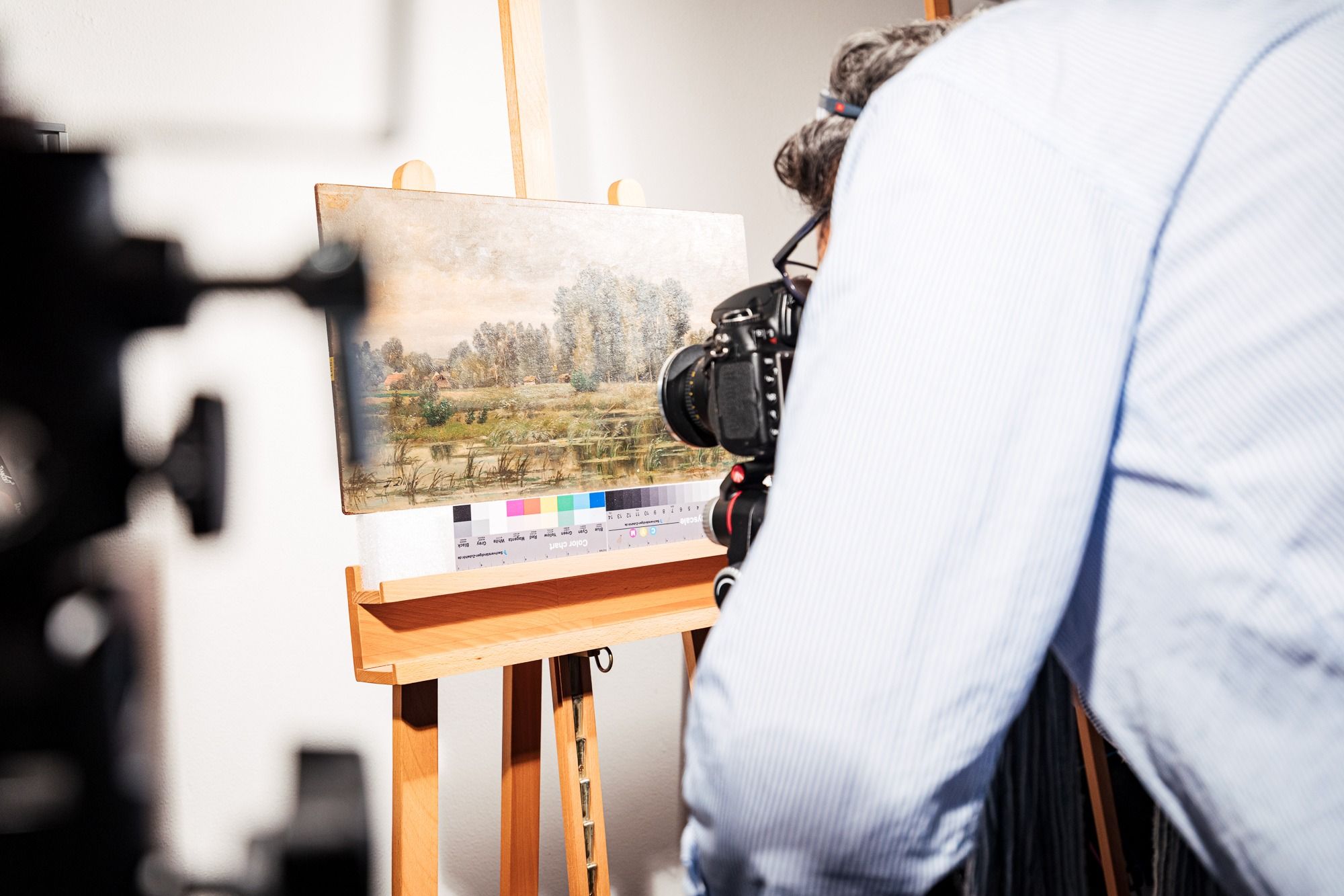
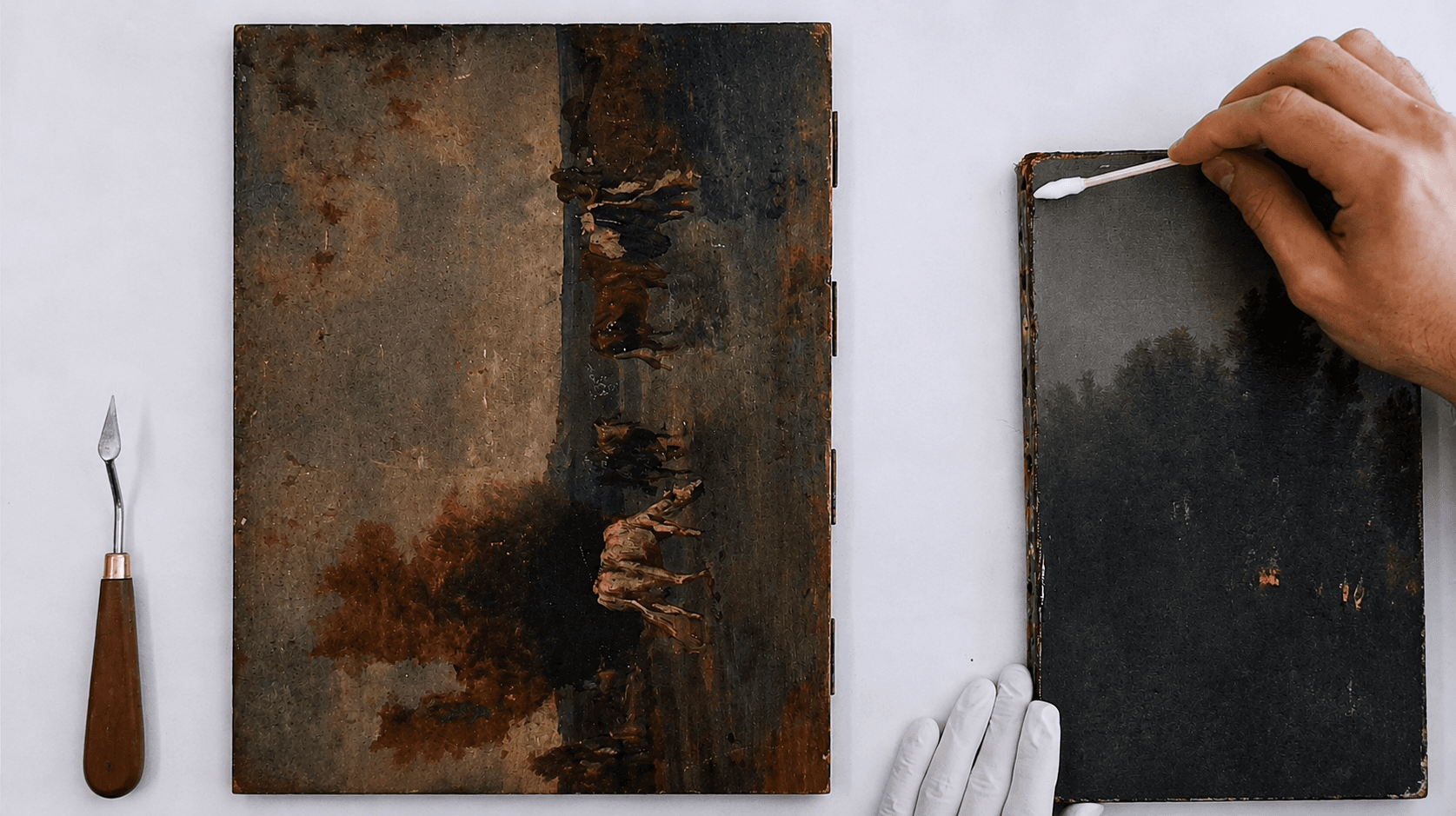
Each artwork tells a unique story and deserves to be preserved with the utmost care. Ageing, environmental influences, structural damage, paint loss or previous restoration can affect its condition over time. Professional artwork restoration helps preserve authenticity, structural integrity and long-term value. Guided by internationally recognised conservation principles, every intervention is carried out with precision, minimal invasiveness and full documentation.
Types of Restoration Services :
- Painting Restoration
- Surface cleaning and varnish removal
- Structural consolidation
- Filling of paint losses
- Pictorial reintegration
- Protective conservation treatments
Frame Restoration
- Cleaning and conservation
- Structural repairs and consolidation
- Reconstruction of missing decorative elements
- Traditional gilding and finishing
- Protective treatments for long-term preservation
Storage
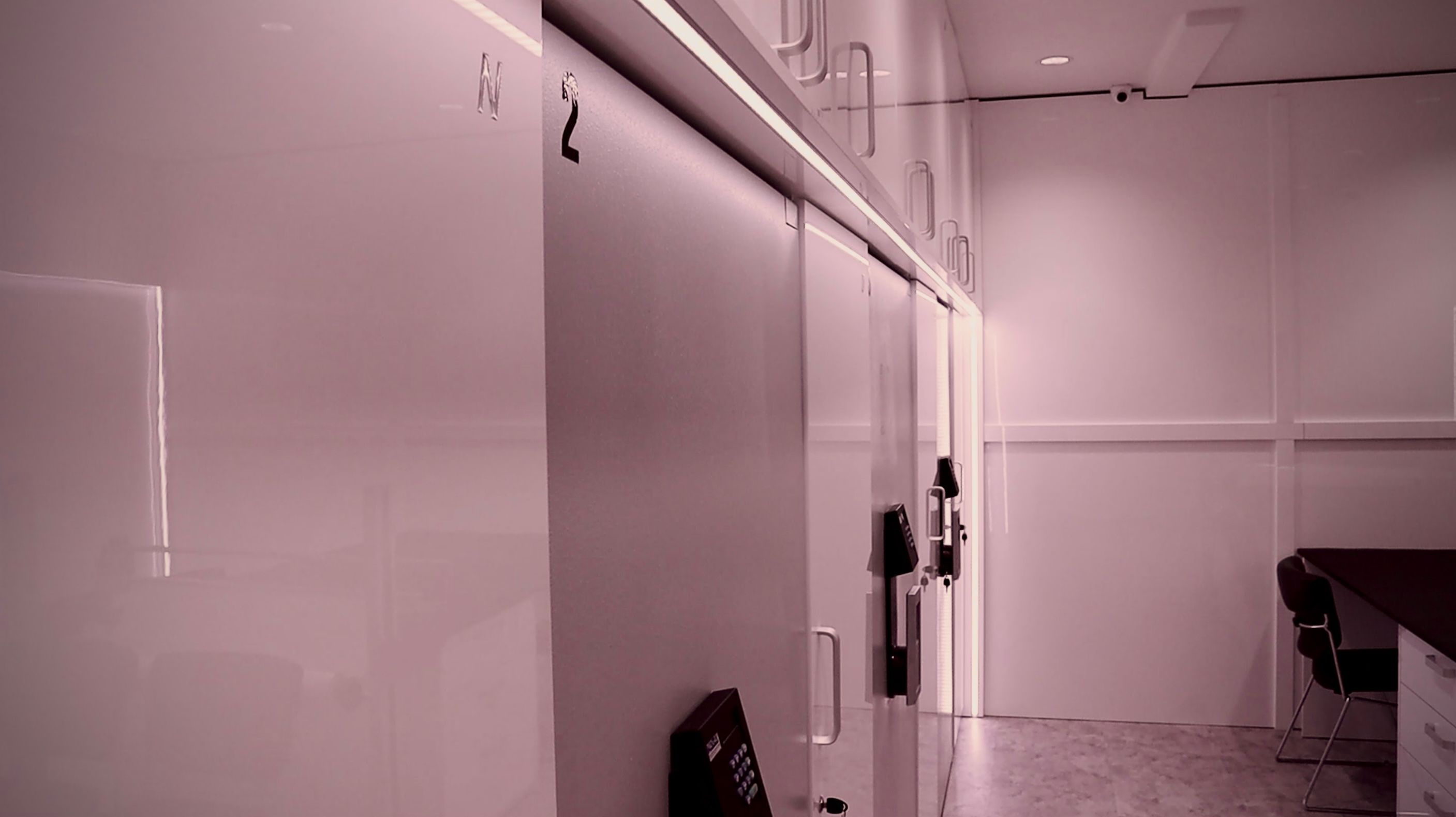
Security and protection are our top priority when it comes to storing artworks. Our secure storage solutions are designed to accommodate a wide range of artworks, ensuring optimal protection through carefully controlled climatic conditions, continuous security monitoring and professional handling. Every artwork is individually documented, assigned a dedicated location and safeguarded in an organised, high-security environment, with access available by prior arrangement.
- 24/7 alarm monitoring
- Immediate security response
- Long-standing security partnership
- Climate-controlled storage
- Efficient access and full traceability
Logistics
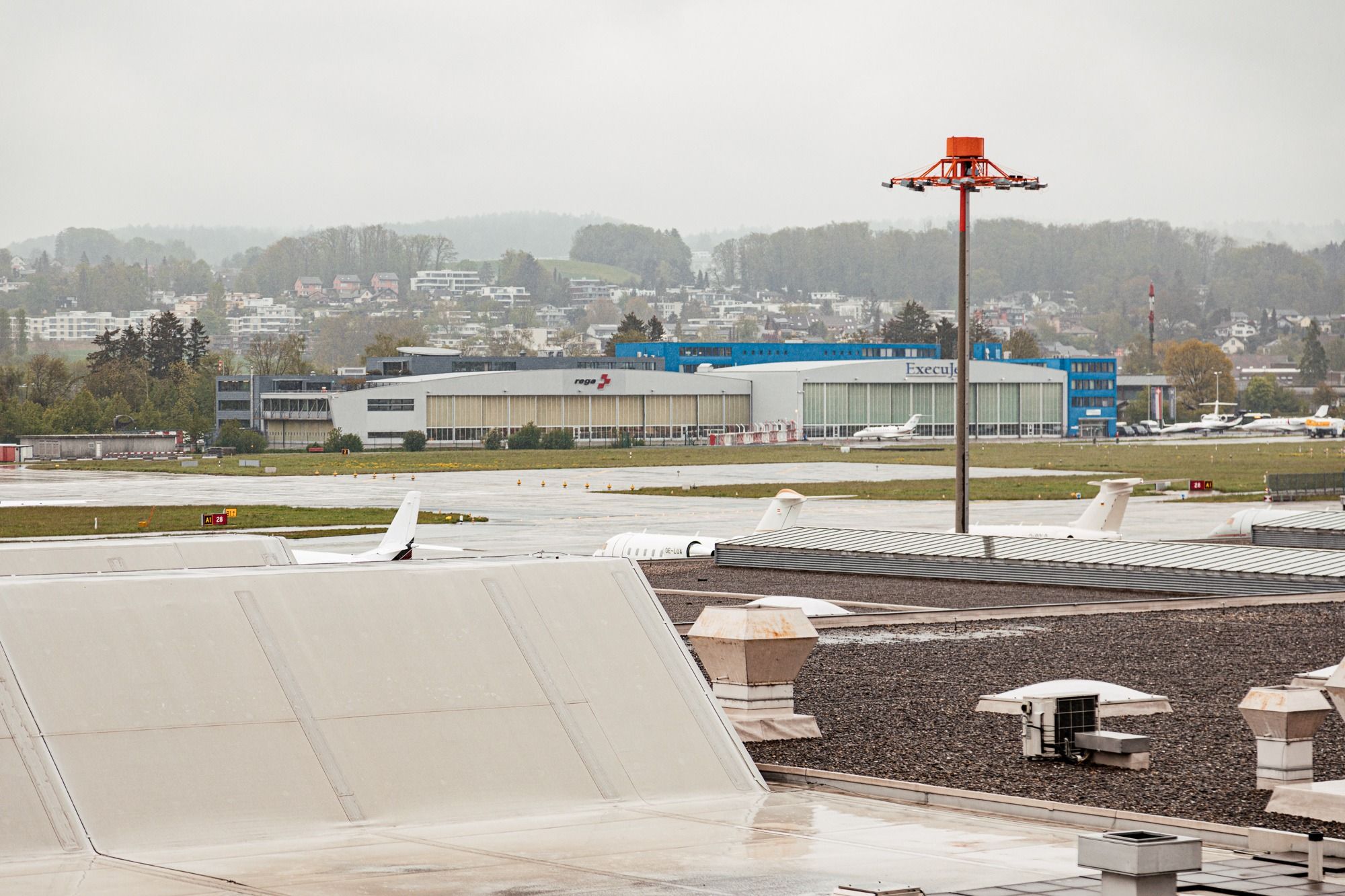
Art travels only as safely as the hands that guide it. We coordinate the secure national and international transport of artworks through our trusted network of specialised fine art logistics partners. Every shipment is individually planned, professionally handled and protected with appropriate transport insurance, ensuring that each artwork reaches its destination safely and under the highest standards of care.
- Transport insurance
- Professional art crating
- Condition and conservation based transport
- Complete transport coordination
- Collection relocation services
- Reliable and efficient delivery
Advisor
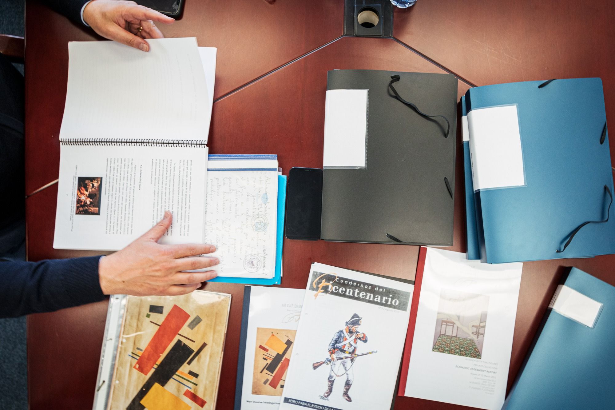
The international art market is complex – we lend clarity, built on experience, strategic insight and trusted guidance. Whether acquiring, selling or evaluating an artwork, we provide independent advice tailored to the individual needs of private collectors, institutions and or professionals. Our objective is to secure the best possible outcome while providing a seamless and confidential experience.
- Guidance on an artwork's significance and market position
- Strategic sales and acquisition support
- International network
- Negotiation assistance
- Transparent client representation
- Access to technical and scientific expertise
Sale & Acquisition
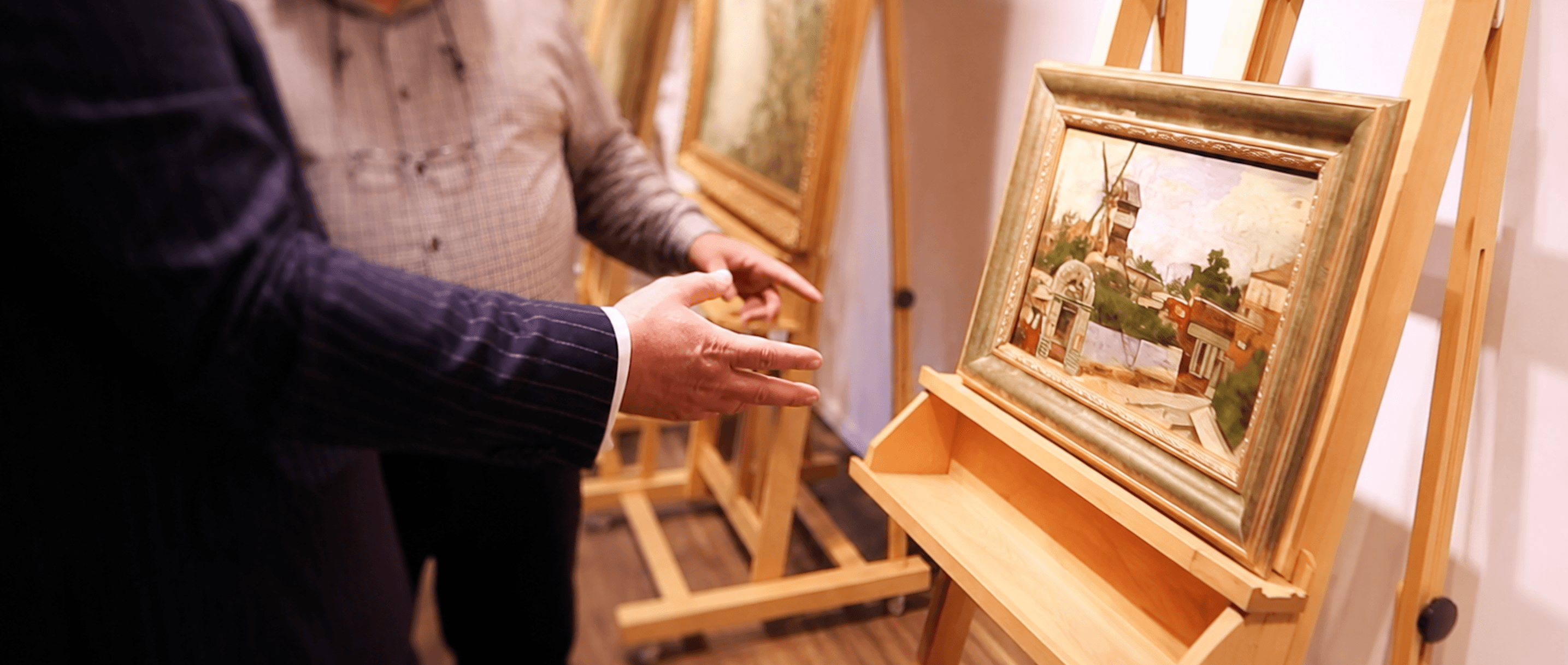
Whether acquiring exceptional artworks for our collection or placing important works with the right collectors and institutions, we approach every transaction with expertise, discretion and a deep understanding of the international art market. Our objective is to strike a fair balance between all parties involved, never losing sight of the true value of the artwork.
- Tailored approach to every transaction
- Viewings by appointment
- Expert-accompanied viewings upon request
- Comprehensive artwork documentation
- Transparency through authenticity, provenance and examination records
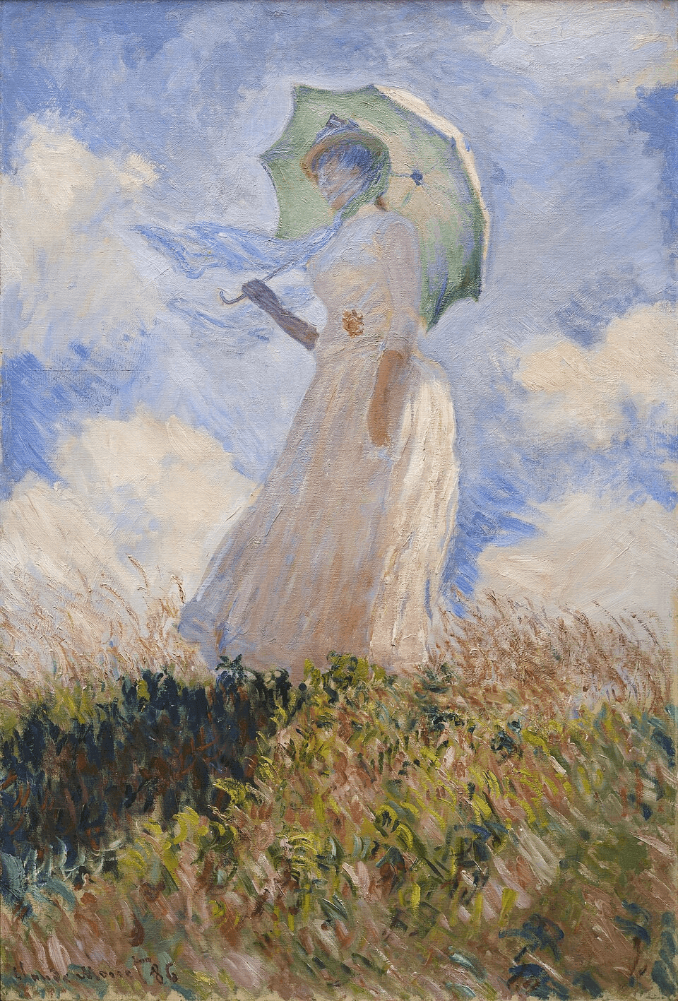
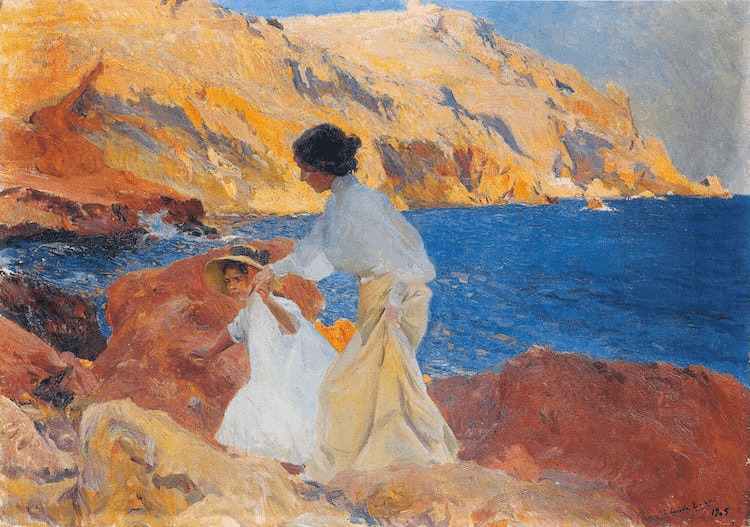
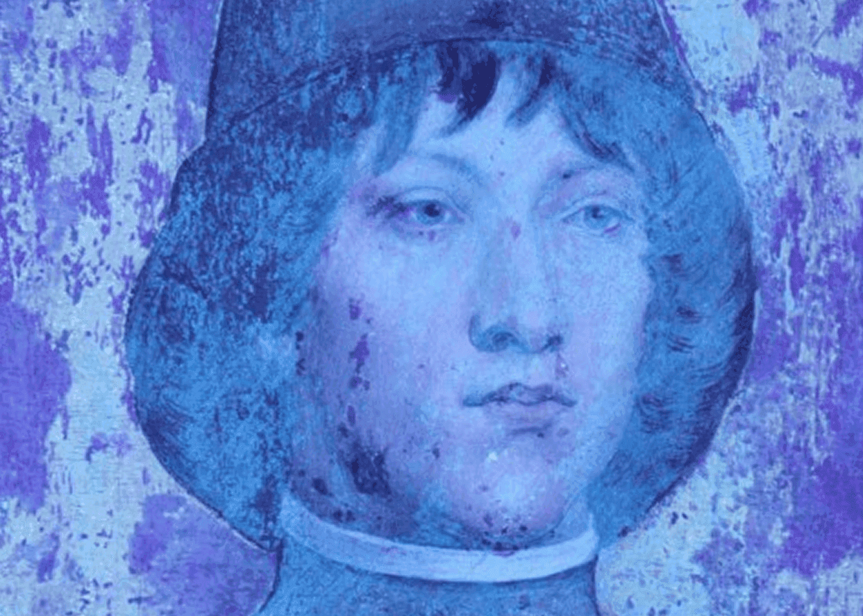
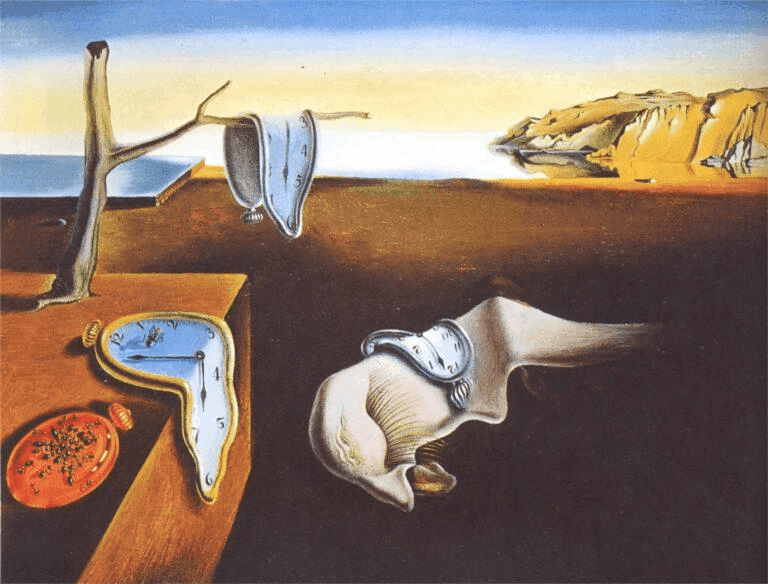
Non-Destructive Investigation Methodology
Technical Photography
Technical photography uses different wavelengths of light and radiation to reveal characteristics of an artwork that may remain invisible under normal viewing conditions. By examining the same area under visible (VIS), ultraviolet (UV) and infrared (IR) radiation, valuable information about the surface, underlying structures, materials and previous interventions can be obtained.
Microscopy
Microscopy enables the detailed examination of an artwork’s surface beyond what is visible to the naked eye. By magnifying fine structural and pictorial features, it provides valuable information about materials, technique, condition and characteristics of execution.
Multispectral Imaging
Multispectral imaging examines an artwork across different regions of the electromagnetic spectrum, including visible, ultraviolet and infrared radiation. By capturing variations in the response of materials across these spectral regions, it can reveal information about pictorial materials and underlying features that may remain invisible under conventional observation.
XSpecam
XpeCAM® is an integrated, portable multispectral imaging system specifically developed by Xpectraltek for the non-invasive documentation, analysis, and conservation of cultural heritage and artworks.
System Overview
The system integrates proven instrumentation and allows you to look beyond the visible spectrum to analyse the layers of artworks, map pigments and safely investigate the history of restorations.
Artificial Intelligence False Colour (AIUVFC & AIIRFC)
Our analytical approach combines multispectral imaging with artificial intelligence to extend the interpretation of false-colour data. A system based on multiple classifiers analyses the acquired images and supports the mapping of pigments across the artwork’s surface, transforming complex imaging data into detailed visual information.
Spectroscopy
Spectroscopic techniques provide analytical information about the materials present within an artwork by examining their interaction with electromagnetic radiation. These methods support the identification and characterisation of pigments and other materials, providing valuable insight into an artwork’s material composition.
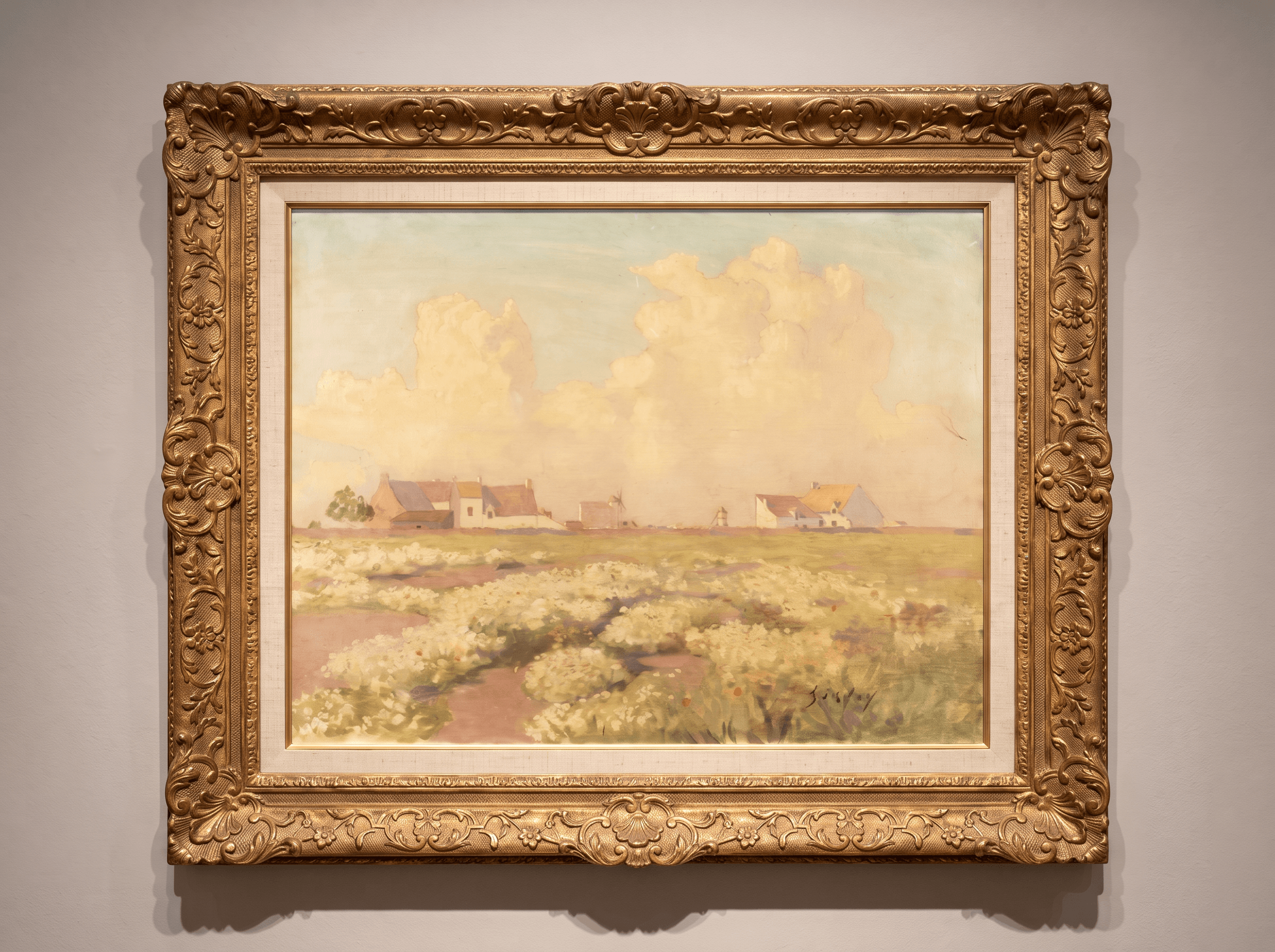
High-resolution photography under controlled visible light provides a precise record of an artwork’s appearance, colour and surface characteristics. It forms an essential reference for documentation, comparison and further technical investigation.
Raking light illuminates the artwork from an oblique angle, emphasising its surface topography. It can reveal impasto, incisions, deformations and subtle structural irregularities, as well as condition phenomena such as lifting paint layers.
By illuminating a painting from behind, transmitted light provides information about the structure and varying transparency of its layers. It can reveal retouching, paint losses, craquelure and other features that may not be clearly visible from the surface.
High-resolution detail photography documents selected areas of an artwork under controlled visible light. It enables the precise examination of features such as signatures, brushwork, surface characteristics, previous interventions and areas of particular interest, providing detailed visual documentation for further technical assessment.
Macrophotography captures highly detailed areas of the artwork at an enlarged scale. It enables close examination of brushwork, craquelure, surface texture and other characteristics relevant to technical and comparative assessment.
Microscopic examination allows the surface of an artwork to be studied at magnifications beyond normal visual observation. Characteristics such as craquelure, brushwork, signatures and paint structures can be examined in detail and evaluated in relation to the technique and presumed period of the artwork.
Microscopy under UV-induced visible fluorescence provides an enhanced view of materials and surface features according to their fluorescence response. It can support the differentiation of materials and the identification of alterations or later interventions at microscopic scale.
Transmitted infrared light can reveal underlying features such as preparatory drawings, underpainting and pentimenti that may not be visible under normal observation. These insights provide valuable information about the construction of the composition and the artist’s working process.
Ultraviolet-induced fluorescence highlights differences in the optical behaviour of materials. It is particularly valuable for detecting retouching, later interventions, varnishes and selected organic or inorganic materials, helping distinguish different phases within an artwork.
Near-infrared photography records differences in the reflection and absorption of infrared radiation across the pictorial surface. These variations can help distinguish materials with different infrared responses and provide additional information about preparatory elements beneath the visible surface.
Ultraviolet False Colour (UVFC) and Infrared False Colour (IRFC) combine UV and infrared imaging with visible-light photography to reveal differences in pictorial materials that are not apparent under conventional observation. The resulting images support pigment characterisation and are further analysed using an AI-based system of five classifiers, enabling detailed pigment mapping across the entire surface of the artwork.
Raman spectroscopy provides molecular information about materials through their interaction with monochromatic light. In the examination of artworks, it is particularly valuable for the identification and characterisation of pigments present within the pictorial surface.
X-ray fluorescence is a non-destructive analytical technique used to determine the elemental composition of materials. By identifying characteristic chemical elements, XRF provides important information for pigment characterisation and the technical investigation of an artwork.
FORS is a non-invasive spectroscopic technique used for the characterisation of pigments, dyes and alteration products, as well as the investigation of colour and colour variations. Portable instrumentation enables measurements to be performed directly on the artwork without sampling.
OUR EXPERTS